This video is available for instant download licensing here https://www.alilamedicalmedia.com/-/galleries/narrated-videos-by-topics/ekgecg/-/medias/4f63a6a7-94c6-463e-8c85-7117e48f5d23-atrial-fibrillation-narrated-animation ©Alila Medical Media. All rights reserved. Support us on Patreon and get FREE downloads and other great rewards: patreon.com/AlilaMedicalMedia All images/videos by Alila Medical Media are for information purposes ONLY and are NOT intended to replace professional medical advice, diagnosis or treatment. Always seek the advice of a qualified healthcare provider with any questions you may have regarding a medical condition. Atrial fibrillation is the most common type of cardiac arrhythmia. In a healthy heart, the sinoatrial node or SA node initiates all electrical impulses in the atria. In atrial fibrillation, electrical impulses are initiated randomly from many other sites called ectopic sites in and around the atria, commonly near the roots of pulmonary veins. These un-synchronized, chaotic electrical signals cause the atria to quiver or fibrillate rather than contract. Although the atrial rate during atrial fibrillation can be extremely high, most of the electrical impulses do not pass through the atrioventricular – the AV – node to the ventricles. This is due to refractory properties of the cells of the AV node. Those do come through are irregular. Ventricular rate or heart rate is therefore irregular and can range from slow – less than 60 – to rapid -more than 100 – beats per minute. On an ECG (EKG), atrial fibrillation is characterized by absence of P-waves and irregular narrow QRS complexes. Reminder: P-wave represents electrical activity of the SA node that is now obscured by activities of multiple ectopic sites. The baseline may appear undulating or totally flat depending on the number of ectopic sites in the atria. In general, larger number of ectopic sites results in flatter baseline. As the atria do not function properly, the heart puts out less blood, and heart failure may occur. The most common complication of atrial fibrillation, however, is the formation of blood clots in the atria. As the atria do not empty completely into the ventricles, the blood may stagnate inside the atria and blood clots may form. These clots may then pass into the bloodstream, get stuck in small arteries and block them. When a blood clot blocks an artery in the brain, a stroke may result.
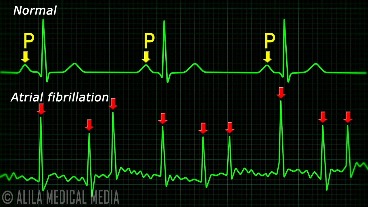
Atrial Fibrillation Anatomy, ECG and Stroke, Animation.
You Might Also Like
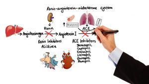
Pharmacology – HYPERTENSION & ANTIHYPERTENSIVES (MADE EASY)

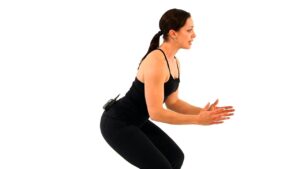
Minute Body | WILD OUTDOORS – Jump Squats

Sporting house Meaning

Why do our bodies need protein?

Vegetarian sources of Omega 3
Muscle Building Workout & Squats Video – 22

Orthopedic Physiotherapy Video – 8
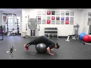
Achieve PT PE | Exercise of the Week: Swiss Ball Reverse Hyperextension
BCAA (Branched Chain Amino Acids)
How To: Side Oblique Crunch

Supplements For Beginners | Complete Supplement Guide For Beginners
How to Do a Hammer Curl | Female Bodybuilding
What Is Type 1 Diabetes? | 2 Minute Guide | Diabetes UK
What is Diabetes?
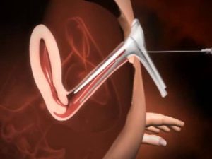
What is IUI treatment for Pregnancy
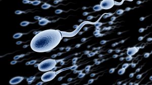
Frequency of Surgical Sperm Retrieval Procedure-Dr. Sathya Balasubramanyam of Cloudnine Hospitals

World’s Best Multivitamins at CHEMIST SHOP | Cheapest | Guaranteed Results
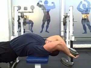
Dumbbell Pullovers A Forgotten Chest Exercise
Spor Salonunda Yapılan Hatalar 18 – Barbell Lying Triceps Extension Nasıl Yapılır

Insulin Does NOT Build Muscle | #RXRants | RXMuscle.com
The endocrine system
Multivitamin Supplement
Muscle Definition – Does Your Training Matter? 8 minute explanation
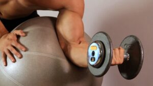
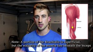
How to Do a Swiss Ball Preacher Curl | Arm Workout

5 Components of Fitness

Uses of Cheapest Vitamin B Complex with B12 NEUROBION FORTE

Free Paleo Meal Plan + Shopping List for weight loss

Easy High Protein Bodybuilding Breakfast
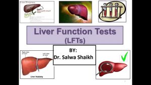
Liver Function Test (LFT) explained by Dr. Salwa Shaikh
How to Do Reverse Hypers with a Swiss Ball
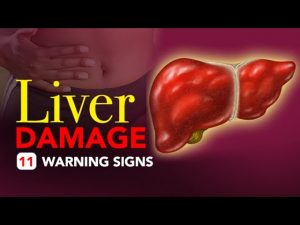
Liver Damage – 11 Warning Signs

What should one have during severe Diarrhea? – Ms. Sushma Jaiswal
Orthopedic Physiotherapy Video – 7
Anatomy on USMLE — What’s High Yield?
Pulley Curl-3

5 Quick Yoga Poses For THYROID Problems & Disorders

Puberty and The Hormones Involved | Physiology | Biology | FuseSchool

Drug and Medicine
If You Eat 2 Eggs at Breakfast For a Month, This is What Happens to Your Body

OneTouch SelectSimple Demo
Step Up-3