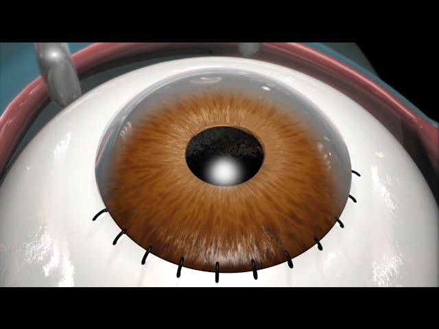
UPDATE 2011: A newer version of this animation is now available! You can watch it here: https://youtu.be/icYLMmENk_c http://www.nucleushealth.com/ – This 3D medical animation depicts the phacoemulsification and extracapsular removal of a cataract (cloudy lens), and the placement of an artificial lens. The lens of the eye is responsible for focusing images onto the back of the eye. It is normally transparent. As a normal part of aging, the lens begins to cloud and causes a gradual, painless loss in vision. Cataract removal is most often performed to better examine the back of the eye when monitoring for damage from certain diseases such as diabetes or glaucoma and to improve vision. There are two main types of cataract removal. The large majority of cataract surgeries are performed using the phacoemulsification technique. During the phacoemulsification technique an ultrasound probe breaks the cloudy lens into tiny fragments. The fragments are vacuumed out through a tiny incision. An intraocular lens implant is then inserted to replace the natural lens that was removed. Because the incision is tiny, stitches are often not necessary and visual improvement is usually noted relatively soon after surgery. During the extracapsular technique the cataract is removed as one entire piece. This requires a larger incision and stitches. An intraocular lens implant (artificial lens) is inserted to replace the natural lens that was removed. Recovery is usually slower, due to the larger incision. The stitches sometimes need to be removed, which is usually done in the office. After both procedures, the surgeon usually places a patch over the eye. ANCE00193
Laser Surgeries Video – 2
You Might Also Like
How to do a Donkey Kick Exercise
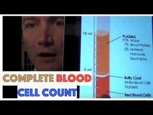
Complete Blood Cell Count

Intermittent Fasting & Fasting Video – 29
How to Do a Side Crunch | Ab Workout
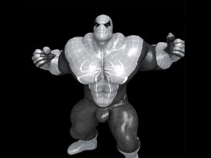
Spiderman Muscle Growth Animation 3

Peck Deck Fly | Chest Exercise #2 | Fitness With Sangram Chougule

Anemia – Causes, Symptoms, Treatments & More…

What is Fish Oil? Omega-3 Benefits & Side Effects Review by Guru Mann
Of Anatomy and Physiology (2013 – 3D Animation)
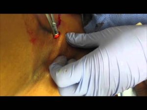
Abscess Basics: Part 2a

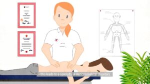
Branches of Physiotherapy Video – 8
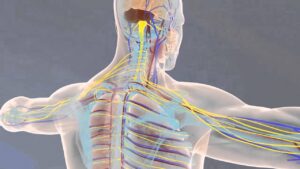
Neurology Video – 1
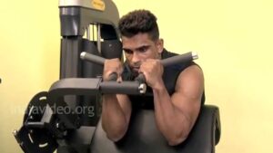
Body Building – Biceps Exercise Guide – Preacher Hammer Curls

Preparation for a BodyBuilding Competition – 20 WEEKS OUT! World Champion Explains

Cure Cirrhosis With This Recipe – Homeveda Shorts

Modified Tricep Dips Demo

Relactation and induced lactation | Breastfeeding

HGH, Growth Hormones & Plant Hormones Video – 25

How to Do a Dumbbell Biceps Curl | Arm Workout

First Aid Video – 4
Leg Raises-2
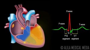
Cardiac Conduction System and Understanding ECG, Animation.

Testosterone & Androgenic Effects Video – 35

Top 10 fruits to eat while Breastfeeding
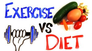
Exercise vs Diet

Vitamin B Complex – What and Why?
Branches of Physiotherapy Video – 17

Fat Loss, Weight Loss Video – 27

KUWTK | Kris Jenner Interferes With Pregnant Khloé’s Workout | E!

Physiotherapy in Obstetrics Video – 6
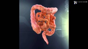
Human Internal Organs 3D Model From CreativeCrash.com

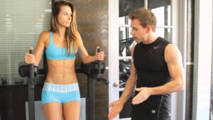
How to warm up before exercise

Developmental Psychology Video – 3

Anaerobic – Medical Definition

Static plank exercise: isometric workout for static strength

5 BCAA Side Effects

Sugar Free, Low Sugar Video – 28

World’s Best Fish Oil – Omega 3 at CHEMIST SHOP | Cheapest | Guaranteed Results

Why do you feel cold when you have fever?
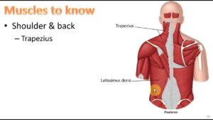
Anatomy Ch 9 – Muscular System

Types of Jaundice in Newborn