Structure of Bone|Anatomy of Bone|3D Animation|Biology It is important for bones to be strong to support our body weight and in some cases provide protection such as the skull and ribs. However, they must also be light enough to make movement possible. A long bone consists of several sections: Diaphysis: This is the long central shaft Epiphysis: Forms the larger rounded ends of long bones Metaphysis: Area betweent the diaphysis and epiphysis at both ends of the bone Epiphyseal Plates: Plates of cartilage, also known as growth plates which allow the long bones to grow in length during childhood. Once we stop growing, between 18 and 25 years of age the cartilage plates stop producing cartilage cells and are gradually replaced by bone. Covering the ends of bones, where they form a joint with another bone, is a layer of hyaline cartiage. This is a firm but elastic type of cartilage which provides shock absorbtion to the joint and has no neural or vascular supply. Bone Anatomy If you were to cut a cross-section through a bone, you would first come across a thin layer of dense connective tissue known as Periosteum. This can be divided into two layers, an outer ‘fibrous layer’ containing mainly fibroblasts and an inner ‘cambium layer’, containing progenitor cells which develop into osteoblasts (the cells responsible for bone formation). The periosteum provides a good blood supply to the bone and a point for muscular attachment. Formation and remodelling of bone Bone formation is an essential process in the development of the human body. It starts during the development of the foetus, and continues throughout childhood and adolescence as the skeleton grows. Bone remodelling meanwhile is a life-long process, consisting of resorption (the breaking down of old bone) and ossification (formation of new bone), and is key to shaping the skeleton and to the repair of bone fractures. There are three types of cell present in bone that are of particular interest – osteoblasts, osteocytes and osteoclasts, which are respectively responsible for the production, maintenance and resorption of bone. Osteoblasts Mononucleated “bone-forming” cells found near the surface of bones. They are responsible for making osteoid, which consists mainly of collagen. The osteoblasts then secrete alkaline phosphatase to create sites for calcium and phosphate deposition, which allows crystals of bone mineral to grow at these sites. The osteoid becomes mineralised, thus forming bone. Osteocytes These are osteoblasts that are no longer on the surface of the bone, but are instead found in lacunae between the lamellae in bone. Their main role is homeostasis – maintaining the correct oxygen and mineral levels in the bone. Osteoclasts Multinucleated cells responsible for bone resorption. They travel to specific sites on the surface of bone and secrete acid phosphatase, which unfixes the calcium in mineralised bone to break it down. During foetal development there are two mechanisms for creating bone tissue: Endochondral ossification Intramembranous ossification Intramembranous ossification occurs in the formation of flat bones such as those in the skull, and will not be covered further here. More information can be found through the Going Further page. Endochondral ossification This involves bone growth from an underlying cartilage model, and is seen in the formation and growth of long bones such as the femur. The initial step involves the development of a cartilage model, which has the rough shape of the bone being formed. In the middle of the shaft is the primary ossification centre, where osteoblasts lay down osteoid on the shaft to form a bone collar. The osteoid calcifies, and blood vessels grow into cavities within the matrix. Osteoblasts then use the calcified matrix as a support structure to lay down more osteoid and form trabeculae within the bone. Meanwhile osteoclasts break down spongy bone to create the medullary cavity, which contains bone marrow.
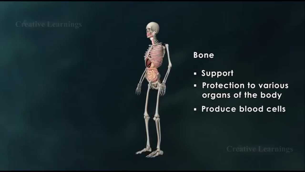
Structure of Bone|Anatomy of Bone|3D Animation|Biology
You Might Also Like

HOW TO LOSE WEIGHT FAST 10Kg in 10 Days | 900 Calorie Egg Diet By Versatile Vicky
HOW TO DRAW HUMAN INTERNAL ORGANS

Bodybuilding Nutrition, Diet Recipes & Workout – 35
Gynecomastia (Male breast) surgery- Dr Pranav Kumar- Patna, Bihar, India
Glutamine Supplements Explained in 60 Seconds – Should You Take It?
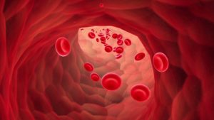
Anaemia
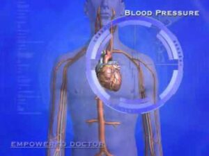
Blood Pressure Explained
Epidemiology Video – 1
Supplements: Truth About Creatine Risks
Endurance

Three Types Of Stretching

How Much Water Should You Drink Per Day?
ENDOCRINE SYSTEM: HYPOTHALAMUS AND CONTROL OF THE PITUITARY
Stroke – Causes, Symptoms and Treatment Options
Pregnancy Exercises Video – 6

Law 3: Train to Lose | 10 Laws of Muscle-Building

Stretch All Deltoid Heads!
Milk Nutrition & Processing Video – 2
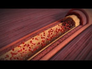
About Your Heart Attack | Nucleus Health
Scripps Health: Menopause Signs, Symptoms and Treatment
What would happen if you didn’t drink water? – Mia Nacamulli

Latissimus Dorsi Bent Over Row-9

Diet & Nutrition : How to Gain Weight
METFORMIN MECHANISM OF ACTION MADE SIMPLE *ANIMATED*
Reversing Type 2 Diabetes Naturally: 3 Inexpensive Foods You Should Know About
What Is Circuit Training? | Gym Workout
Initial Symptoms That Your Blood Pressure Is Dangerously High. (1 OF 5 DO NOT KNOW THEY HAVE IT)!

Geriatric Psychiatry Video – 1
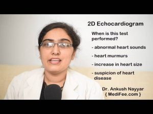
2D Echocardiogram – Testing for Heart Health
Personal Trainer/ Gym Instructor Video – 6

Mushroom Nutrition Video – 2

Weight Loss Drugs, Orlistat: the good and bad
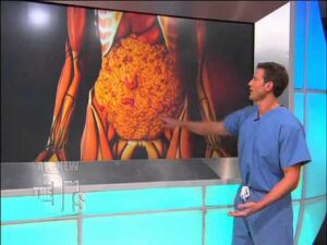
What Visceral Fat Does To The Body (The Doctors)
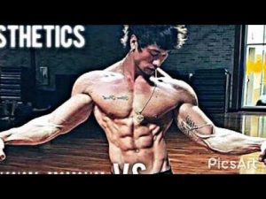
Jon Skywalker “The Definition Of Aesthetics” | Fitness & Bodybuilding Motivation
Hypertension & Blood Preesure

Suicide Psychology/ Psychiatry Video – 2

6 Worst Exercises That WILL Get You Injured

Propranolol
Bench Dips for Big Triceps, How to Get Big Triceps
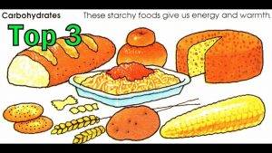
Top 3 Carb Food For Lean Muscle Growth

Surya Namaskar Video – 4