UPDATE 2011: A newer version of this animation is now available! You can watch it here: https://youtu.be/icYLMmENk_c http://www.nucleushealth.com/ – This 3D medical animation depicts the phacoemulsification and extracapsular removal of a cataract (cloudy lens), and the placement of an artificial lens. The lens of the eye is responsible for focusing images onto the back of the eye. It is normally transparent. As a normal part of aging, the lens begins to cloud and causes a gradual, painless loss in vision. Cataract removal is most often performed to better examine the back of the eye when monitoring for damage from certain diseases such as diabetes or glaucoma and to improve vision. There are two main types of cataract removal. The large majority of cataract surgeries are performed using the phacoemulsification technique. During the phacoemulsification technique an ultrasound probe breaks the cloudy lens into tiny fragments. The fragments are vacuumed out through a tiny incision. An intraocular lens implant is then inserted to replace the natural lens that was removed. Because the incision is tiny, stitches are often not necessary and visual improvement is usually noted relatively soon after surgery. During the extracapsular technique the cataract is removed as one entire piece. This requires a larger incision and stitches. An intraocular lens implant (artificial lens) is inserted to replace the natural lens that was removed. Recovery is usually slower, due to the larger incision. The stitches sometimes need to be removed, which is usually done in the office. After both procedures, the surgeon usually places a patch over the eye. ANCE00193
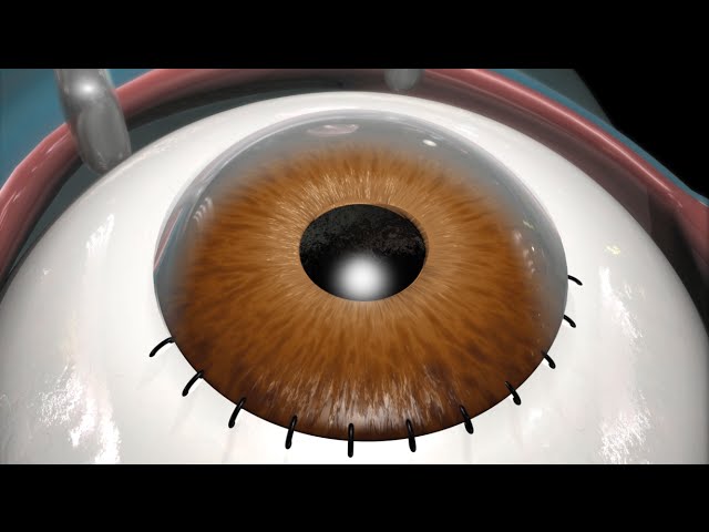
Laser Surgeries Video – 2
- Post author:
- Post published:May 16, 2021
- Post comments:0 Comments
You Might Also Like

Supplement Spotlight Silymarin

K-Cold Anti Cold Tablet Full Review

How to Dumbbell Squat | Mike Hildebrandt

Diabetes symptoms in men | early diabetes symptoms in men

What causes Diarrhea? + more videos | #aumsum #kids #science #education #children

Importance of Lipid Profile Test

Nitroglycerin: Explosive Heart Medication

Water Nutrition Video – 1

Brain 101 | National Geographic

What is cardiovascular exercise — Definition of cardiovascular endurance

Canoeing Video – 4

Best Fat Burning Workout (HOW I STAY LEAN!)
Workouts for Women to Lose Belly Fat at HOME
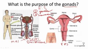
8.2.7 Gonads – Structure and Function

These Are The Symptoms and Signs You May Have a Blood Clot in Your Leg
Organ Transplantation Surgeries Video – 4

What causes headache vomiting and diarrhea ? | Health Channel

Side Crunch With Weight-2

Intermittent Fasting & Fasting Video – 11

Post Workout Drink | GET RIPPED Male & Female FITNESS MODEL Program by Guru Mann

No Gym Full Body Workout

Home Exercise for Spinal Cord Injury: Back Extension

How To: Dumbbell Side Lateral Raise

B1 Homeostasis Blood Glucose and Diabetes EDEXCEL

Diseases Caused by Malnutrition – SCURVY, RICKETS, BERIBERI, PELLAGRA

What To Eat Before A Cross Country Race
Gynecomastia

When His Penis Won’t Stand Up: Causes of Erectile Dysfunction | Quint Fit

Fasting + Post Meal Blood Sugar Readings

Muscular Strength Asanas Video – 6

How to Do Basic Sitting Stretches | Taekwondo Training

Human Body, Body Building Muscle Building Anatomy Physiology Video – 46
Latissimus Dorsi Bent Over Row-5
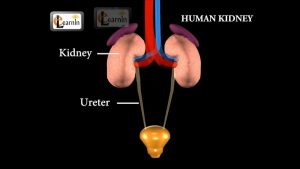
Kidney – Excretory System – Biology

What is Aerobic Exercise- Cardio and aerobics workouts

The Best Way To Use Creatine!! Get Amazing Results Fast

Sports Medicine Video – 4
Hormone Levels

Proper Form For Dumbbell Flyes

Preparation for a BodyBuilding Competition – 20 WEEKS OUT! World Champion Explains

Benefits of a Creatine Supplement – Know Your Supps

