Structure of Bone|Anatomy of Bone|3D Animation|Biology It is important for bones to be strong to support our body weight and in some cases provide protection such as the skull and ribs. However, they must also be light enough to make movement possible. A long bone consists of several sections: Diaphysis: This is the long central shaft Epiphysis: Forms the larger rounded ends of long bones Metaphysis: Area betweent the diaphysis and epiphysis at both ends of the bone Epiphyseal Plates: Plates of cartilage, also known as growth plates which allow the long bones to grow in length during childhood. Once we stop growing, between 18 and 25 years of age the cartilage plates stop producing cartilage cells and are gradually replaced by bone. Covering the ends of bones, where they form a joint with another bone, is a layer of hyaline cartiage. This is a firm but elastic type of cartilage which provides shock absorbtion to the joint and has no neural or vascular supply. Bone Anatomy If you were to cut a cross-section through a bone, you would first come across a thin layer of dense connective tissue known as Periosteum. This can be divided into two layers, an outer ‘fibrous layer’ containing mainly fibroblasts and an inner ‘cambium layer’, containing progenitor cells which develop into osteoblasts (the cells responsible for bone formation). The periosteum provides a good blood supply to the bone and a point for muscular attachment. Formation and remodelling of bone Bone formation is an essential process in the development of the human body. It starts during the development of the foetus, and continues throughout childhood and adolescence as the skeleton grows. Bone remodelling meanwhile is a life-long process, consisting of resorption (the breaking down of old bone) and ossification (formation of new bone), and is key to shaping the skeleton and to the repair of bone fractures. There are three types of cell present in bone that are of particular interest – osteoblasts, osteocytes and osteoclasts, which are respectively responsible for the production, maintenance and resorption of bone. Osteoblasts Mononucleated “bone-forming” cells found near the surface of bones. They are responsible for making osteoid, which consists mainly of collagen. The osteoblasts then secrete alkaline phosphatase to create sites for calcium and phosphate deposition, which allows crystals of bone mineral to grow at these sites. The osteoid becomes mineralised, thus forming bone. Osteocytes These are osteoblasts that are no longer on the surface of the bone, but are instead found in lacunae between the lamellae in bone. Their main role is homeostasis – maintaining the correct oxygen and mineral levels in the bone. Osteoclasts Multinucleated cells responsible for bone resorption. They travel to specific sites on the surface of bone and secrete acid phosphatase, which unfixes the calcium in mineralised bone to break it down. During foetal development there are two mechanisms for creating bone tissue: Endochondral ossification Intramembranous ossification Intramembranous ossification occurs in the formation of flat bones such as those in the skull, and will not be covered further here. More information can be found through the Going Further page. Endochondral ossification This involves bone growth from an underlying cartilage model, and is seen in the formation and growth of long bones such as the femur. The initial step involves the development of a cartilage model, which has the rough shape of the bone being formed. In the middle of the shaft is the primary ossification centre, where osteoblasts lay down osteoid on the shaft to form a bone collar. The osteoid calcifies, and blood vessels grow into cavities within the matrix. Osteoblasts then use the calcified matrix as a support structure to lay down more osteoid and form trabeculae within the bone. Meanwhile osteoclasts break down spongy bone to create the medullary cavity, which contains bone marrow.
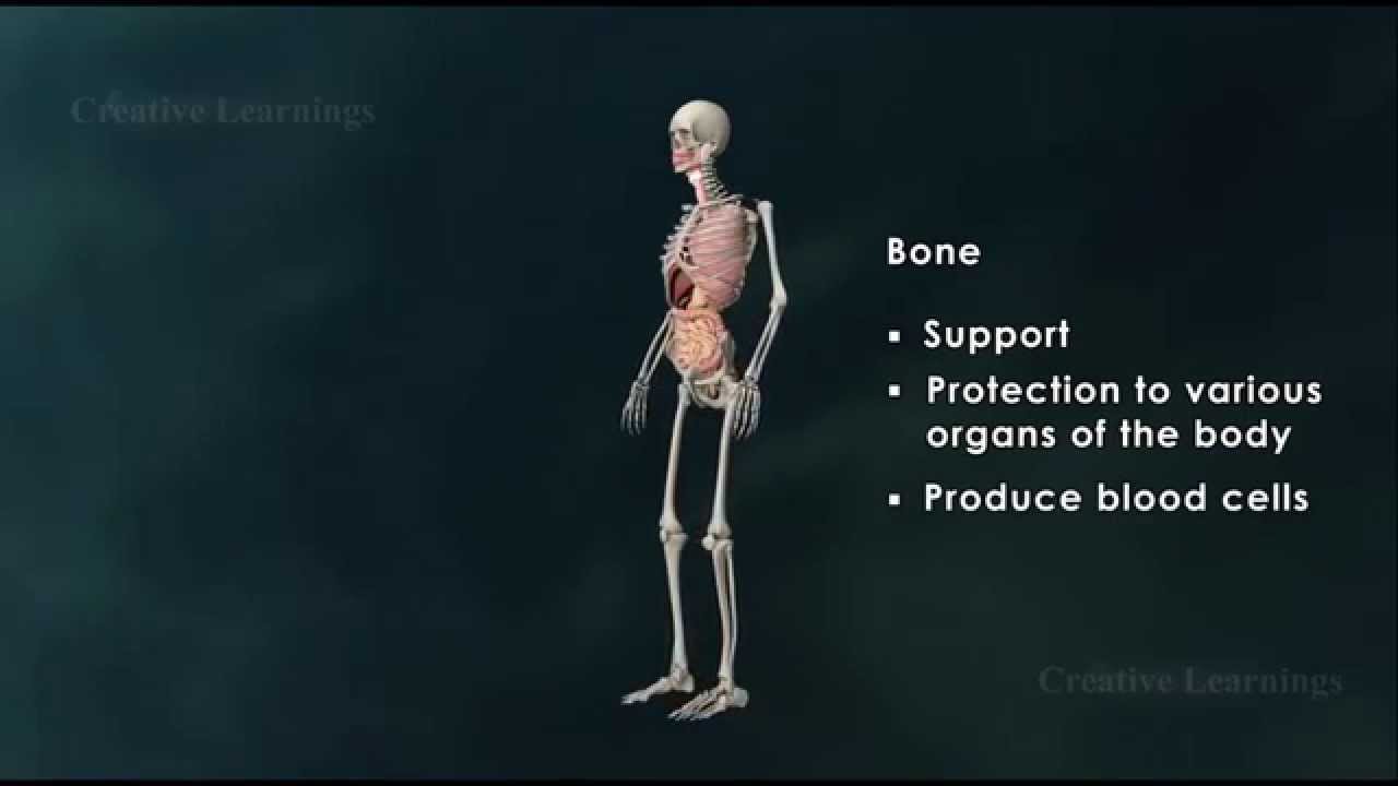
Structure of Bone|Anatomy of Bone|3D Animation|Biology
You Might Also Like
Pharmacodynamics
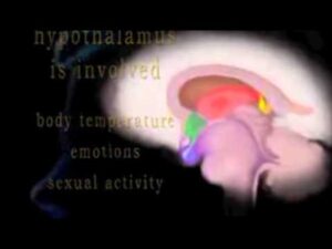
Endocrine System
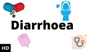
What is Diarrhoea? Causes, Signs and Symptoms, Diagnosis and Treatment.
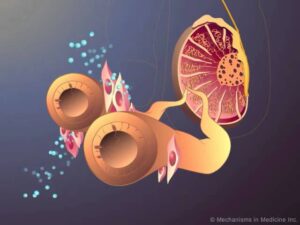
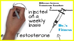
Testosterone Production
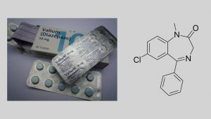
Diazepam (Valium): What You Need To Know
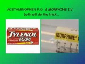
Medication Onset of Action & Peak Effect

Canoeing Video – 3
Fat Burning Process
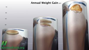
Meat and Weight Gain in the PANACEA Study

Handball Video – 1
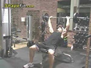
Incline Bench Press-8
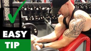
EASY TIP – How to Preacher Curl for Big Gains
Surya Namaskar Video – 3

ANTIOXIDANTS: THE BASICS | Nutrition 101 Ep. 4
What is Jaundice?Causes, Signs and symptoms, Diagnosis and treatment

One Direction – Perfect (Official Video)
Home Cholesterol Test (Total & HDL) Demonstration
Fat Loss, Weight Loss Video – 18
Lee Priest Gives T3 T4 Advice
Hyperextension With ball-9

Are BCAAs A Waste Of Money???

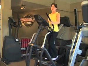
Muscle Building Workout & Squats Video – 8

Spa Resort Video – 2
LOWER ABS UNLEASHED – 3 Exercises! (V-CUT Abs)
Hair Transplant 3d Animation
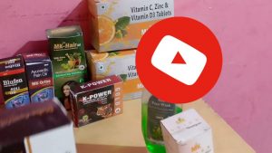
so herbal medicine product video for this YouTube

Top 5 Fruits That Burn Belly Fat
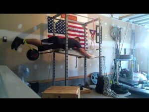
Homemade reverse hyper machine
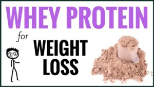
How to Use Whey Protein for Weight Loss
8. Secondary Action – 12 Principles of Animation
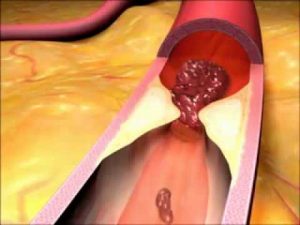
Heart attack in 3d animation

Intermittent Fasting & Fasting Video – 8
Rear Deltoid-9
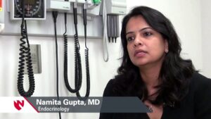
Endocrinology Video – 1

Beginners Guide To Getting FIT
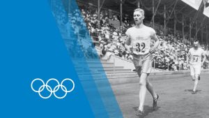
Modern Pentathlon Video – 3

Pre & Post Workout Nutrition Guide – How To DIY TV
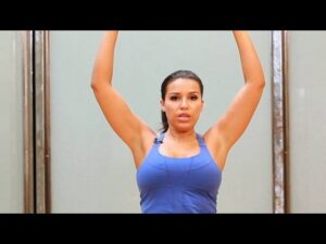
How to Do an Overhead Press | Female Bodybuilding
Testosterone & Androgenic Effects Video – 50
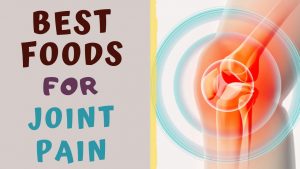
DIET FOR JOINT PAIN – Best Foods for people with Arthralgia
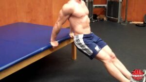
Triceps Dips-5