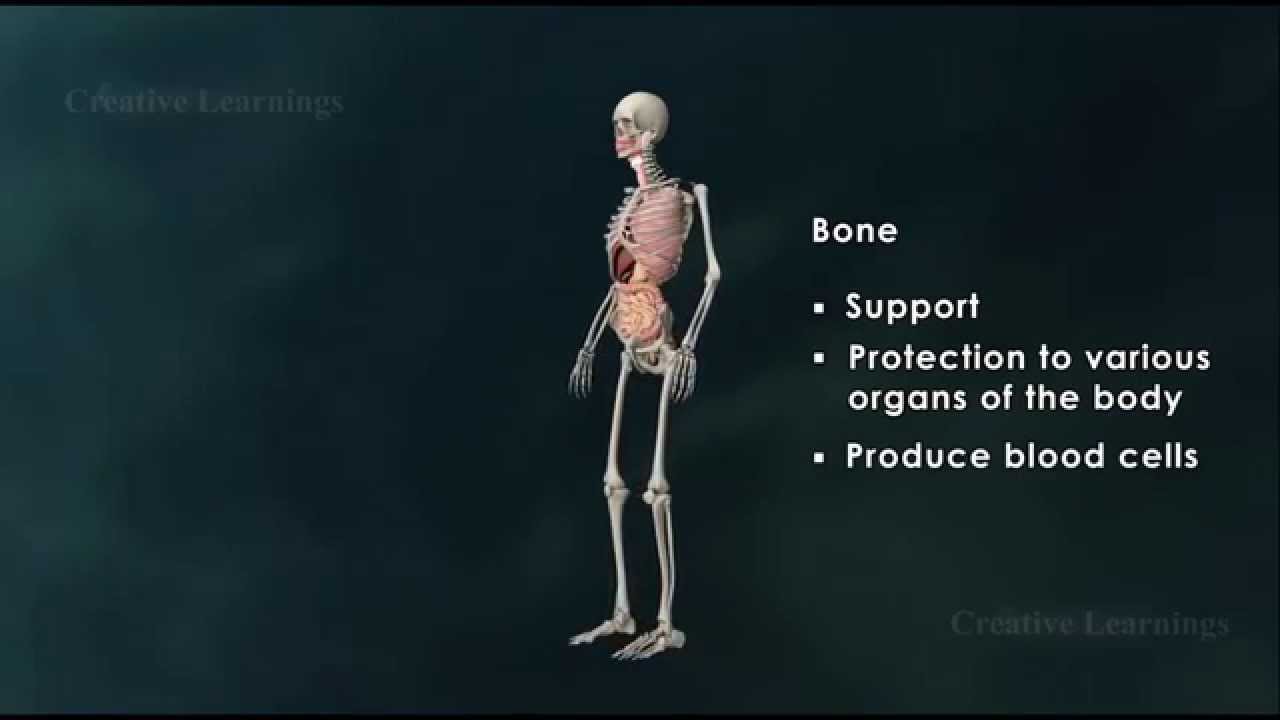
Structure of Bone|Anatomy of Bone|3D Animation|Biology It is important for bones to be strong to support our body weight and in some cases provide protection such as the skull and ribs. However, they must also be light enough to make movement possible. A long bone consists of several sections: Diaphysis: This is the long central shaft Epiphysis: Forms the larger rounded ends of long bones Metaphysis: Area betweent the diaphysis and epiphysis at both ends of the bone Epiphyseal Plates: Plates of cartilage, also known as growth plates which allow the long bones to grow in length during childhood. Once we stop growing, between 18 and 25 years of age the cartilage plates stop producing cartilage cells and are gradually replaced by bone. Covering the ends of bones, where they form a joint with another bone, is a layer of hyaline cartiage. This is a firm but elastic type of cartilage which provides shock absorbtion to the joint and has no neural or vascular supply. Bone Anatomy If you were to cut a cross-section through a bone, you would first come across a thin layer of dense connective tissue known as Periosteum. This can be divided into two layers, an outer ‘fibrous layer’ containing mainly fibroblasts and an inner ‘cambium layer’, containing progenitor cells which develop into osteoblasts (the cells responsible for bone formation). The periosteum provides a good blood supply to the bone and a point for muscular attachment. Formation and remodelling of bone Bone formation is an essential process in the development of the human body. It starts during the development of the foetus, and continues throughout childhood and adolescence as the skeleton grows. Bone remodelling meanwhile is a life-long process, consisting of resorption (the breaking down of old bone) and ossification (formation of new bone), and is key to shaping the skeleton and to the repair of bone fractures. There are three types of cell present in bone that are of particular interest – osteoblasts, osteocytes and osteoclasts, which are respectively responsible for the production, maintenance and resorption of bone. Osteoblasts Mononucleated “bone-forming” cells found near the surface of bones. They are responsible for making osteoid, which consists mainly of collagen. The osteoblasts then secrete alkaline phosphatase to create sites for calcium and phosphate deposition, which allows crystals of bone mineral to grow at these sites. The osteoid becomes mineralised, thus forming bone. Osteocytes These are osteoblasts that are no longer on the surface of the bone, but are instead found in lacunae between the lamellae in bone. Their main role is homeostasis – maintaining the correct oxygen and mineral levels in the bone. Osteoclasts Multinucleated cells responsible for bone resorption. They travel to specific sites on the surface of bone and secrete acid phosphatase, which unfixes the calcium in mineralised bone to break it down. During foetal development there are two mechanisms for creating bone tissue: Endochondral ossification Intramembranous ossification Intramembranous ossification occurs in the formation of flat bones such as those in the skull, and will not be covered further here. More information can be found through the Going Further page. Endochondral ossification This involves bone growth from an underlying cartilage model, and is seen in the formation and growth of long bones such as the femur. The initial step involves the development of a cartilage model, which has the rough shape of the bone being formed. In the middle of the shaft is the primary ossification centre, where osteoblasts lay down osteoid on the shaft to form a bone collar. The osteoid calcifies, and blood vessels grow into cavities within the matrix. Osteoblasts then use the calcified matrix as a support structure to lay down more osteoid and form trabeculae within the bone. Meanwhile osteoclasts break down spongy bone to create the medullary cavity, which contains bone marrow.
Structure of Bone|Anatomy of Bone|3D Animation|Biology
You Might Also Like
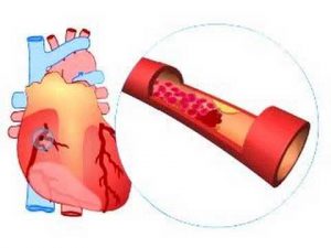
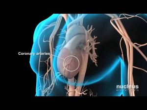
What causes a heart attack?

TIPS | Rujuta Diwekar| Food for Breastfeeding Indian Moms ( Episode 2)
Psychiatry & Psychology
Incline Dumbbell Row
Penile Prosthesis Animation Sequence
How To Use Protein Powder: Simple Step-By-Step Guide

Dermatology & Skin Diseases : Accutane Benefits

8 Foods to Avoid During Pregnancy II Pregnancy Diet Plan II Diet in First Trimester to Third Trimest
How your adrenal hormones affect thyroid and sex hormones
Preacher Curl On Cable Machine
Thyroid Disease Explained in Telugu | Patient Education I MIC

Nephrology Video – 2
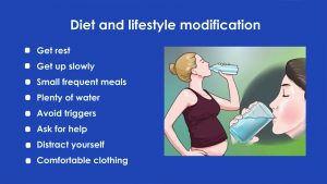
How to Deal With Morning Sickness During Pregnancy? | Dr. Jyoti Kala
Cholesterol Blood Test

Preparing your Body for Pregnancy | Kaiser Permanente
Testosterone & Androgenic Effects Video – 7
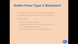
Study 2 – Type 2 Diabetes with Metformin
The Difference Between Sperm & Semen – Episode 20
How to warm up before exercise
How A DEXA Scan Works
Diet Plan for 6 Pack Abs (STEP BY STEP!)
Biceps Cable Preacher Curl Exercises Guide! Live Health Club
How to Do a Seated Calf Raise | Female Bodybuilding
Tennis Video – 2
The scientific truth about steroids
How To: Glute Bridge
Glucagon
Geriatrics Video – 1

Muscleblaze(BCAA)and(L-Arginine)rs.638 Pre workout i by FROM AMAZON | bodybuilding supplements
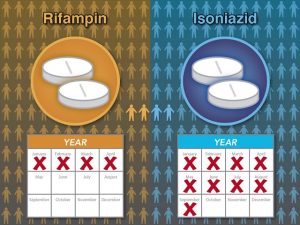
Treating Latent Tuberculosis
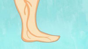
Pregnancy swelling (edema)
StairMaster HIIT Programming Solutions
Bowflex® Bodyweight Workout | Three-Minute Perfect Plank
Universal Nutrition Gain Fast – Powerful Anabolic Weight Gain Formula

25 Different Squat Exercises
![Read more about the article Drug Induced Renal Failure – Causes, Symptoms & Stages [Doctor Interview]](https://videos.drmaheshkumar.com/wp-content/uploads/2021/05/Drug-Induced-Renal-Failure-Causes-Symptoms-Stages-Doctor-Interview-300x169.jpg)
Drug Induced Renal Failure – Causes, Symptoms & Stages [Doctor Interview]
Static Quadriceps Exercise, Correct technique (Isometric Quads)

Alli weight loss pills
Diet & Exercise For Pregnant Women I 3
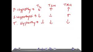
Medical Video Lecture: How to diagnose thyroid diseases quickly with T3,T4, TSH, TRH
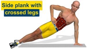
Six pack abs: Side plank with crossed legs