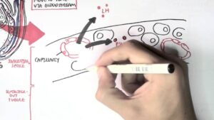
“How to Perform a Transthoracic Echocardiographic Study Volume 1: Transducer Position and Anatomy” is an instructional video, offered by ASE, and can be used for professional lectures and offers an interactive section for flexible presentations. The video includes an overview of relevant cardiac anatomy, a step by step presentation of all Transducer Positions, and the sequential transducer movements to acquire standard echo images needed to complete a Transthoracic Echocardiographic Study. The cardiac anatomy section visualizes relationships of the heart, ribs, axis of the heart, planes of the heart and aorta, chambers, valves and annuli, and blood flow through the heart. The transducer position menu allows the user to view the anatomical landmarks and the transducer positions for left parasternal, apical, subcostal, suprasternal and right parasternal image acquisition. The views and imaging menu allows the user to navigate from a particular transducer position menu such as left parasternal long to specific images acquired within that window such as PLAX high depth, PLAX low depth, and PLAX Zoom of the Aortic and Mitral Valves. Each animated loop shows a finder image in the upper left that depicts the transducer position, an illustration of the sectional anatomy of the heart on the left, and the echo image with labels on the right.
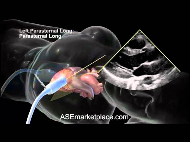
How to do a Basic Transthoracic Echocardiogram: Transducer Position and Anatomy
You Might Also Like
Wrestling Video – 2

What to Eat Before and After Workout at Gym | bodybuilding tips
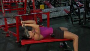
The Ab Bench Back Extension : Training & Body Sculpting
Flat Bench Fly-2

First Aid Video – 4

This Is What Happens To Your Body After Drinking Red Bull
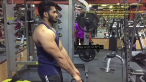
Close Grip Triceps Extension-7

Raw Pressery | All Fruits. No Additives.

Warning: Dangerous Fish Oil Omega 3 Side Effects

Anemia In Children – Causes, Symptoms and Treatment
BUILD SHIRT STRECHING BACK WITH UPPER TRAPEZIUS-ROPE SHRUGS WORKOUT.

Foods to give when child has diarrhoea
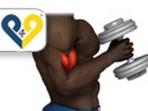
Alternating hammer curls (standing with dumbbells)
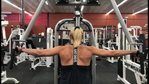
Shrugs-2
The Human Body Video – 5
What is Fitness World Gym’s?

Foods Nutrition Video – 2

3 Simple Home Remedies To Cure WEAKNESS IN BODY(Asthenia)
Human Body, Body Building Muscle Building Anatomy Physiology Video – 38

HGH, Growth Hormones & Plant Hormones Video – 44

Extreme sport Meaning

Robotics Surgeries Video – 4
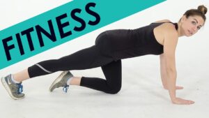
12 Exercises To Change Your Life
Ask a cardiologist: Lipid profile: All you need to know (Expert opinion)
Male Reproductive System – Hormonal Function and Regulation (sperm synthesis and maturation)
ONDANSETRON
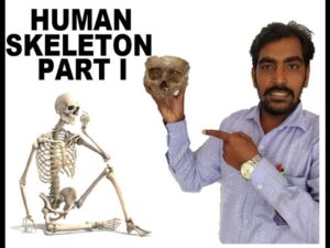
HOW TO REMEMBER HUMAN SKELETON 206 BONES – PART I SKULL
elitefts.com—Mid-Back Extension / Upper Back Shrug
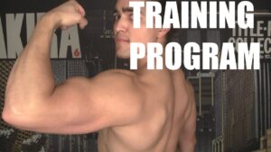
HOW to DESIGN a DAMN GOOD PROGRAM: 4 Key Pillars
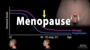
Menopause, Perimenopause, Symptoms and Management, Animation.

CBC test lern video
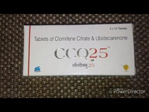
CCQ 25 Tablets Review | Clomifene Citrate & Ubidecarenone Tablets Uses, Side Effects, Precautions
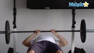
How to Do Lying Barbell Extensions

Complete Blood Count with Normal Values
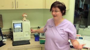
CBC Machine Maintenance
Introduction To Anatomy Physiology: Body Cavities (01:07)

What does isotretinoin mean?
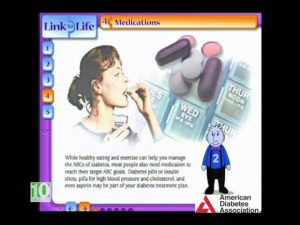
Diabetes Education
Diet Tips: 7 Best Foods To Increase Body Strength
Human Body, Body Building Muscle Building Anatomy Physiology Video – 18

Thyroid gland and hormone synthesis