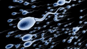
GET LECTURE HANDOUTS and other DOWNLOADABLE CONTENT FROM THIS VIDEO SUPPORT US ON PATREON OR JOIN HERE ON YOUTUBE. https://www.patreon.com/medsimplified The testis is the male gland important for both reproductive and endocrine functions. Initially, it begins as an undifferentiated gonad in the retroperitoneal area. Transcription of the SRY gene (testis-determining factor region) on the Y chromosome ultimately leads to sex differentiation. Without the SRY gene, the gonad would develop into an ovary. As the fetus develops, the functioning testis produces the male hormone testosterone to allow development of male genitalia. Over the last 3 months of gestation, the testis must course its way down from its original retroperitoneal position to its final destination in the scrotum. During its journey it must pass through the peritoneum, abdominal wall via the inguinal canal, and into the scrotal pouch. The testis is a paired, ovoid male reproductive organ that sits in the scrotum, separated from its mate by a scrotal septum. Described by some as being shaped and sized like a large olive or small plum, the average volume of the adult testis is approximately 25 mL. Typically, it measures 3.5-5 cm in length by 2.5-3 cm in both width by 3cm in depth The tunica vaginalis testis (a remnant of the processus vaginalis) envelopes the testis in a double layer, except at the superior and posterior borders where the spermatic cord and epididymis adhere to the testes. The visceral layer of the tunica vaginalis testis is closely applied to the testis, epididymis, and ductus deferens. On the posterolateral surface of the testis, this layer invests a slit-like recess between the body of the epididymis and the testis that is called the sinus of epididymis.[5] The parietal layer of tunica vaginalis is adjacent to the internal spermatic fascia, is more extensive, and extends superiorly into the distal part of the spermatic cord. Deep to the tunica vaginalis, the tunica albuginea is a tough, fibrous outer covering of the testis. On the posterior surface, it is reflected inwardly to form an incomplete vertical septum called the mediastinum testis. The mediastinum testis extends from the superior to near the inferior portion of the gland. It narrows in width as it travels inferiorly. Anteriorly and laterally, numerous imperfect septa are given off, which radiate to the glands surface and are attached to the tunica albuginea. These divide the interior of the testis into numerous, cone-shaped spaces that have a wide base at the gland’s surface and narrow as they converge to the mediastinum. In these spaces, the numerous lobules of glandular structures (the minute but long and highly coiled seminiferous tubules) are housed. The mediastinum supports the ducts and vessels as they pass to and from the glandular substance. The seminiferous tubules are lined with germ cells that produce sperm and nutrient fluid. These tubules empty their contents into a network of anastomosing ducts, which ultimately empties into the epididymis. he epididymis is a comma shaped, elongated structure composed of a single, fine tubular structure estimated up to 6 meters (approximately 20 feet) in length. This tube highly convoluted and tightly compressed (average size of approximately 5 cm) to the point of appearing solid. Located on the posterior border of the testis, it is composed of 3 parts, including the head (caput), body (corpora), and tail (cauda). The epididymal head overhangs the upper pole of the testis, receives the seminal fluid from the ducts of the testis (which pierce the upper portion of the mediastinum), then allows the passage of the sperm into the distal portion of the epididymis. Due to its length, the epididymal duct allows space for storage and maturation of sperm. Progressively tapering in width, the narrow tail continues as the ductus deferens Subscribe here – https://www.youtube.com/channel/UCOmrniWfKi-uCD6Oh6fqhgw Watch Again : https://youtu.be/ImetvbMXgUA -~-~~-~~~-~~-~- CHECK OUT NEWEST VIDEO: “Nucleic acids – DNA and RNA structure ” https://www.youtube.com/watch?v=0lZRAShqft0 -~-~~-~~~-~~-~-
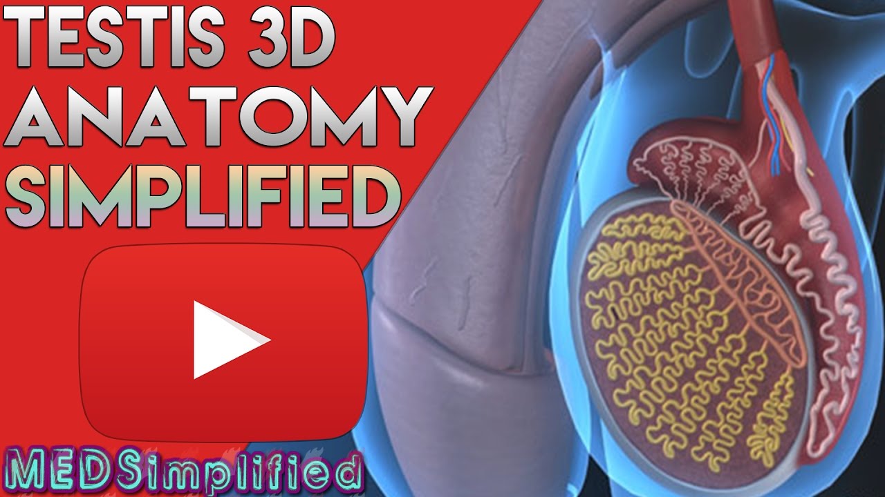
Testis and Epididymis – Male Reproductive Anatomy Part 1
You Might Also Like
Hip Flexor Stretch – Done Correctly

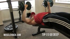
5 MUST-TRY Techniques For Chest Definition

Diabetes Nutrition

Steroids BAD! TRT GOOD! The Effects On Your Heart (Latest Studies)

Sugar Free, Low Sugar Video – 18
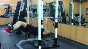
Dumbbell Pullover-3
Frequency of Surgical Sperm Retrieval Procedure-Dr. Sathya Balasubramanyam of Cloudnine Hospitals

Nutrition Advice : What Vitamins Help Arthritis?

Sports Surgeries Video – 1
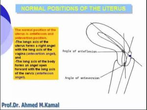
23 Pelvis Normal Positions of Uterus

What is Diabetes Mellitus?
Hair Fall Protection Medicines Biotin
Preservatives Video – 3
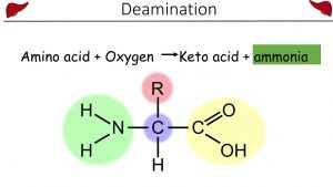
Urea and the Ornithine cycle
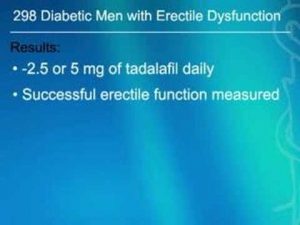
Daily Tadalafil Prevents Erectile Dysfunction in Diabetic
HGH, Growth Hormones & Plant Hormones Video – 43

9 Best TIPS TO GAIN WEIGHT By Dietitian Jyoti Chabria
Estrogen Dominance

ओमेगा 3 क्या होता है और कैसे इस्तेमाल करे | Omega 3 Fish oil Benefits | In 2019 | In Hindi
Latissimus Dorsi Bent Over Row-10

Fat Loss, Weight Loss Video – 29

The Scariest Side Effect of Pregnancy!
Laproscopic Surgeries Video – 1

Pediatric Surgery Video – 2

General Surgery Video – 5

Nutrition education improves diabetes, says study

Wide grip upright rows for side delts
HOW TO DO Hamstring Exercise – Leg Curl with Resistance Bands

Silymarin – Rejuvenate your Liver

Spiking insulin naturally to build muscle and lose fat

FAT LOSS VEGETARIAN Diet Plan for Women (Hindi) | How to Lose Weight Fast 10kgs | Indian Meal Plan

Zenith Nutrition Silymarin Milk Thistle Review (Hindi)
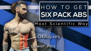
HOW TO TRAIN ABS (Scientific Workout for SIX PACK) | Best Exercises for UPPER, LOWER ABS & OBLIQUES
How the Body Absorbs and Uses Medicine | Merck Manual Consumer Version

The GET RIPPED Chest Workout (MINI ROUTINE MAX GAINS!)
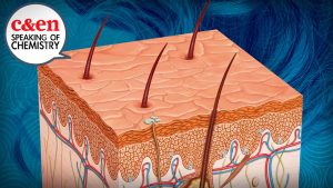
Why Does Your Hair Turn Gray? – Speaking of Chemistry
Science Says Decline Dumbbell Bench Press Is BEST Chest Exercise
Barbell Decline Bench Press – HASfit Lower Chest Exercise Demonstration – Decline Press – Pectoral

Dumbbell Chest Workout (INCOMPLETE WITHOUT THIS!)
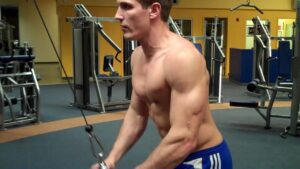
Triceps Pulley Extension-2
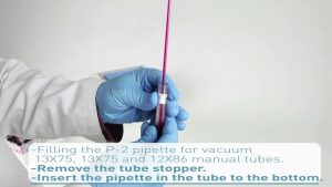
Aquisel – Tubes and pipettes