✪✪✪✪✪ http://www.theaudiopedia.com ✪✪✪✪✪ What is ECHOCARDIOGRAPHY? What does ECHOCARDIOGRAPHY mean? ECOCARDIOGRAPHY meaning. Echocardiogram, often referred to as a cardiac echo or simply an echo, is a sonogram of the heart. (It is not abbreviated, as ECG is an abbreviation for an electrocardiogram.) Echocardiography uses standard two-dimensional, three-dimensional, and Doppler ultrasound to create images of the heart. Echocardiography has become routinely used in the diagnosis, management, and follow-up of patients with any suspected or known heart diseases. It is one of the most widely used diagnostic tests in cardiology. It can provide a wealth of helpful information, including the size and shape of the heart (internal chamber size quantification), pumping capacity, and the location and extent of any tissue damage. An echocardiogram can also give physicians other estimates of heart function such as a calculation of the cardiac output, ejection fraction, and diastolic function (how well the heart relaxes). Echocardiography can help detect cardiomyopathies, such as hypertrophic cardiomyopathy, dilated cardiomyopathy, and many others. The use of Stress Echocardiography may also help determine whether any chest pain or associated symptoms are related to heart disease. The biggest advantage to echocardiography is that it is noninvasive (doesn’t involve breaking the skin or entering body cavities) and has no known risks or side effects. Not only can an echocardiogram create ultrasound images of heart structures, but it can also produce accurate assessment of the blood flowing through the heart by Doppler echocardiography, using pulsed or continuous wave Doppler ultrasound. This allows assessment of both normal and abnormal blood flow through the heart. Color Doppler as well as spectral Doppler is used to visualize any abnormal communications between the left and right side of the heart, any leaking of blood through the valves (valvular regurgitation), and to estimate how well the valves open (or do not open in the case of valvular stenosis). The Doppler technique can also be used for tissue motion and velocity measurement, by Tissue Doppler echocardiography. Echocardiography was also the first ultrasound subspecialty to use intravenous contrast. (See Contrast Echocardiography) Echocardiography is performed by cardiac sonographers, cardiac physiologists (UK) or doctors trained in echocardiography. Recognized as the “Father of Echocardiography”, the Swedish physician Inge Edler (1911-2001), a graduate of Lund University, was the first of his profession to apply in diagnosing cardiac disease ultrasonic pulse echo imaging technique, a technique that the acoustical physicist Floyd Firestone had developed to detect defects in metal castings. In fact, Dr. Edler in 1953 produced the first echocardiographs using an industrial Firestone-Sperry Ultrasonic Reflectoscope. In developing echocardiography, Edler worked with the physicist Carl Hellmuth Hertz, the son of the Nobel laureate Gustav Hertz and grandnephew of Heinrich Rudolph Hertz.
What is ECHOCARDIOGRAPHY? What does ECHOCARDIOGRAPHY mean? ECOCARDIOGRAPHY meaning
You Might Also Like

Baby Cries During Breastfeeding – Reasons and Solutions
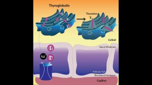
Thyroid Hormone Synthesis

How To Do: Preacher Curl, Good Form vs. Bad Form
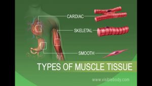
Anatomy and Physiology of Tissues
BCAA Supplement vs Protein Supplement – Know Your Supps – BPI Sports

Antioxidants and Depression

Acupuncture/Acupressure Video – 2

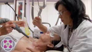
Cardiac surgery Video – 2
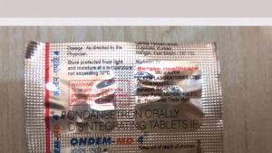
Ondem MD Tablet Review | उल्टी रोकने की दवा। ondansetron medicine |
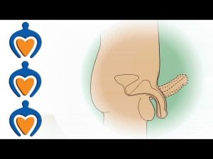
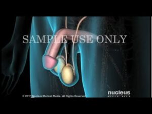
Erectile Dysfunction (ED) – Causes, symptoms and treatment modalities
The Best Exercises for Major Muscle Groups

Antioxidants and Depression
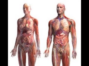
Anatomy and Physiology of Human Body
Dermatology/Skin Surgeries Video – 4

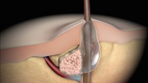
What causes gluteal & perianal abscess? – Dr. Nagaraj B. Puttaswamy

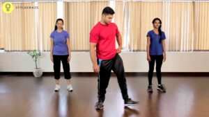
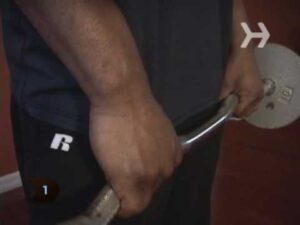
Step Up-4
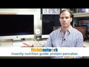
Insanity Nutrition Guide: Protein Pancakes

Love Psychology Video – 1

Pathology Video – 1
30 DAYS SQUAT CHALLENGE : Cool Down Exercises
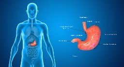
Brain Stroke Can Cause Paralysis Of The Stomach
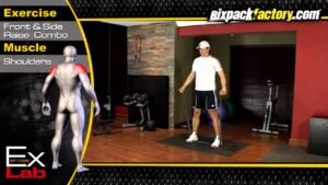
How to Do Rear Deltoid Rows
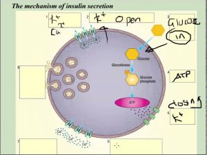
Insulin secretion at cellular level (beta cells) – A2 Science
Lateral Raises-7

Muscle Building Workout & Squats Video – 2

TOP 5 BEST AMINO ACID SUPPLEMENTS OF 2015
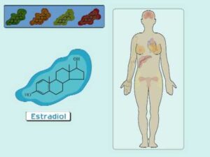
The Estrogen Receptor (I): Hormonal Mechanisms in the Body

Health And Fitness Video – 7

Chances of pregnancy with Gonadotropin and HCG injection – Dr. Sangeeta Gomes

Does masturbation reduce sperm count? | Metromale Clinic & Fertility Center
HGH, Growth Hormones & Plant Hormones Video – 48

Muscle Building Workout & Squats Video -46

Pregnancy Exercises Video – 2

Testosterone & Androgenic Effects Video – 32
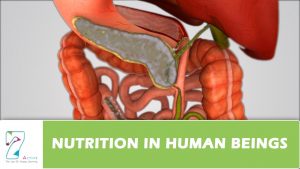
Nutrition in Human Beings
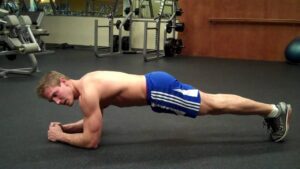
Plank- 2
Acne | Nucleus Health
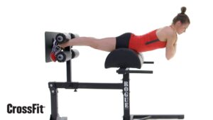
The GHD Back Extension

Omega 3 Fatty Acids के फायदे और नुकसान – Wow, Amway, Healthkart, MuscleBlaze, Healthviva Review

Pulmonology Video – 3

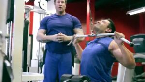
Preacher Hammer Curl – Biceps Exercises