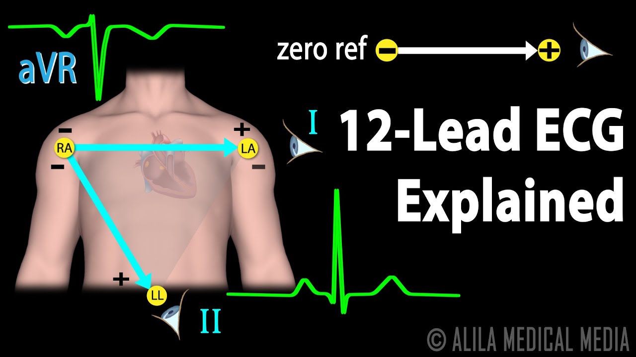
(USMLE topics, cardiology) Understanding the standard 12-lead EKG – Basics of electrocardiography explained. This video is available for instant download licensing here: https://www.alilamedicalmedia.com/-/galleries/narrated-videos-by-topics/ekgecg/-/medias/4d57ce72-0d39-4525-b523-329941b9edcf-12-lead-ecg-explained-narrated-animation ©Alila Medical Media. All rights reserved. Voice by: Sue Stern. Support us on Patreon and get FREE downloads and other great rewards: patreon.com/AlilaMedicalMedia All images/videos by Alila Medical Media are for information purposes ONLY and are NOT intended to replace professional medical advice, diagnosis or treatment. Always seek the advice of a qualified healthcare provider with any questions you may have regarding a medical condition. Electrical activities of the heart can be picked up on the skin via electrodes. An ECG machine records these activities and displays them graphically. The graphs show the heart’s OVERALL electrical potential, or voltage, as it changes over time during a cardiac cycle. The 12 leads of the ECG represent 12 electrical views of the heart from 12 different angles. The conventional 12-lead procedure involves attaching 10 electrodes to the body: one to each limb and six across the chest. There are 6 limb leads and 6 chest leads. The 6 limb leads look at the heart in a vertical plane and are obtained from three electrodes attached to the right arm, left arm, and left leg. The electrode on the right leg is an earth electrode. The measurement of a voltage requires 2 poles: negative and positive. The ECG machine uses the negative pole as zero reference. Thus, the position of the positive pole is the “point of view”, and the line connecting the 2 poles is the “line of sight”. Leads I, II, and III are BI-polar – they measure electrical potential between 2 of the 3 limb electrodes: Lead I represents the voltage between the right arm – negative pole – and the left arm – positive pole, and thus looks at the heart from the left. Lead II sees signal movements between the right arm – negative – and the left leg –positive – forming the INFERIOR LEFT view. Similarly, lead III measures electrical potential between the left arm – negative – and the left leg –positive, looking at the heart from an INFERIOR RIGHT angle. Leads aVR, aVL, and aVF, or “augmented limb leads”, are UNIpolar. They use ONE limb electrode as the positive pole, and take the average of inputs from the OTHER two as the zero reference. Hence, aVR looks at the UPPER RIGHT side of the heart; aVL looks at the UPPER LEFT side of the heart; and aVF looks at the INFERIOR wall of the heart. The chest leads, or precordial leads, view the heart in a HORIZONTAL plane. These are unipolar leads. The corresponding chest electrodes serve as the positive poles. The reference negative value is the same for all chest leads and is calculated as the average of inputs from the three limb electrodes. DE-polarization TOWARD a lead produces a POSITIVE deflection; DE-polarization AWAY from a lead gives a NEGATIVE deflection. The REVERSE is true for RE-polarization. Thus, leads that look at the heart from different angles may have waves pointing in different directions.
12 Lead ECG Explained, Animation
You Might Also Like

Health & Skill Components of Fitness

Bodybuilding Nutrition, Diet Recipes & Workout – 36

Can multivitamins cause any side effects? – Ms. Sushma Jaiswal

Close Grip Bench – HASfit Triceps Exercise Demonstration – Close Bench Triceps Press – Tricep
Acne Protective Medicines Isotretinoin

Intermittent Fasting & Fasting Video – 14

History Of Medicine Video – 2
Liver anatomy and function | Human Anatomy and Physiology video 3D animation | elearnin

RHOMBOIDS & LATS – DB Alternating Bent Over Row

Dumbel 1 arm lying triceps extension
Human Digestive system explained

How to Do Lying Barbell Extensions

Overweight & Obesity Video – 24

Abnormal Psychology Video – 3

How To: Tricep Close Grip Machine Press

Preacher Curl On Cable Machine
Bodybuilding Video – 4
Latissimus Dorsi Bent Over Row-5

Fruits NOT for Pregnant Women
Protein Requirement According To Workout

Pre Workout Drink | LEAN MODE by Guru Mann | Health and Fitness

Health Check: Thyroid Test

… 12 07 2014 – Birmingham – Definition Fitness Centre – Anita CK …

Massage Spa Video – 6

Volleyball Video – 4

2 Main Causes Of Kidney Disease You Must Know

How are MAGGI Noodles made?

Anabolic Steroids – History, Definition, Use & Abuse Video – 2

Insulin & Glucagon
Sports Physiology Video – 3

Nutrition 101 For Students: Carbohydrates

Surgical Debridement Of A Wound – Manipal Hospital
How to Burn Fat Better

How many sets and reps to build muscle | for size, mass, strength

The Truth about BCAAs…

How Much Protein Should You Consume Post-Workout?
Dynamic Stretches to WARM UP Chest Muscles (before you bench!)

Legal Psychology Video – 4

How to Do a Dumbbell Row | Back Workout

How to do Ez Lying Triceps Extensions Correctly | Triceps Exercise

Shoulder Girdle Muscle Group – Kinesiology Quiz