✪✪✪✪✪ http://www.theaudiopedia.com ✪✪✪✪✪ What is ECHOCARDIOGRAPHY? What does ECHOCARDIOGRAPHY mean? ECOCARDIOGRAPHY meaning. Echocardiogram, often referred to as a cardiac echo or simply an echo, is a sonogram of the heart. (It is not abbreviated, as ECG is an abbreviation for an electrocardiogram.) Echocardiography uses standard two-dimensional, three-dimensional, and Doppler ultrasound to create images of the heart. Echocardiography has become routinely used in the diagnosis, management, and follow-up of patients with any suspected or known heart diseases. It is one of the most widely used diagnostic tests in cardiology. It can provide a wealth of helpful information, including the size and shape of the heart (internal chamber size quantification), pumping capacity, and the location and extent of any tissue damage. An echocardiogram can also give physicians other estimates of heart function such as a calculation of the cardiac output, ejection fraction, and diastolic function (how well the heart relaxes). Echocardiography can help detect cardiomyopathies, such as hypertrophic cardiomyopathy, dilated cardiomyopathy, and many others. The use of Stress Echocardiography may also help determine whether any chest pain or associated symptoms are related to heart disease. The biggest advantage to echocardiography is that it is noninvasive (doesn’t involve breaking the skin or entering body cavities) and has no known risks or side effects. Not only can an echocardiogram create ultrasound images of heart structures, but it can also produce accurate assessment of the blood flowing through the heart by Doppler echocardiography, using pulsed or continuous wave Doppler ultrasound. This allows assessment of both normal and abnormal blood flow through the heart. Color Doppler as well as spectral Doppler is used to visualize any abnormal communications between the left and right side of the heart, any leaking of blood through the valves (valvular regurgitation), and to estimate how well the valves open (or do not open in the case of valvular stenosis). The Doppler technique can also be used for tissue motion and velocity measurement, by Tissue Doppler echocardiography. Echocardiography was also the first ultrasound subspecialty to use intravenous contrast. (See Contrast Echocardiography) Echocardiography is performed by cardiac sonographers, cardiac physiologists (UK) or doctors trained in echocardiography. Recognized as the “Father of Echocardiography”, the Swedish physician Inge Edler (1911-2001), a graduate of Lund University, was the first of his profession to apply in diagnosing cardiac disease ultrasonic pulse echo imaging technique, a technique that the acoustical physicist Floyd Firestone had developed to detect defects in metal castings. In fact, Dr. Edler in 1953 produced the first echocardiographs using an industrial Firestone-Sperry Ultrasonic Reflectoscope. In developing echocardiography, Edler worked with the physicist Carl Hellmuth Hertz, the son of the Nobel laureate Gustav Hertz and grandnephew of Heinrich Rudolph Hertz.
What is ECHOCARDIOGRAPHY? What does ECHOCARDIOGRAPHY mean? ECOCARDIOGRAPHY meaning
You Might Also Like
How to check man sperm count for fertility at home

Orthopedic Physiotherapy Video – 8

Is BCCA Good For Your Body? | BeerBiceps BCAA 101
Blood Sugar Testing

Dr Ramakrishna tells about the diet in Arthritis | Online Health Tips
Endometrial Biopsy
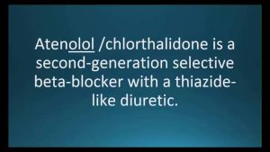
How to pronounce atenolol / chlorthalidone (Tenoretic) (Memorizing Pharmacology Flashcard)
How To: Tricep Close Grip Machine Press

Logan Paul Workout (GYM COMPILATION)

bodybulding
Gene Cloning in Plain English
Onco Surgery Video – 2

What am I eating to BUILD MUSCLES? Full day of eating

Hygiene And House Keeping Video – 1
Q35 elliptical machine from Octane Fitness
What is a Complete Blood Count?
Carbonic Anhydrase Inhibitors EASY mnemonic | Pharmacology Mnemonics

Spa Associations Video – 4
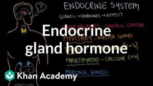
Endocrine gland hormone review | Endocrine system physiology | NCLEX-RN | Khan Academy

Aerobic Exercise Jogging
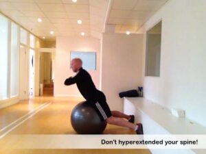
Swiss Ball Back Extension – Medium
This system burns body fat to ash!

Spa Products Video – 1

Aromatic Oils Video – 4

Friday WOD @ DEFINITION FITNESS GYM

Insomnia Video – 2
Decline Bench Press-2
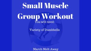
Small Muscle Group Workout/Food is Fuel?
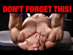
Dumbbell Chest Workout (INCOMPLETE WITHOUT THIS!)
Blood Pooling in Hand

Minoxidil before after – Part 1

Handball Video – 4

Spa Business Video – 4
How Vaccines Work in the Human Body Animation – Immune System Response to Vaccination Video

Side Effects of eating RAW EGGS | Info by Guru Mann
Home Chest Workout (UPPER, MID, LOWER CHEST)
![Read more about the article Bodybuilding Diet – Drinking Water Helps Build Muscle And Lose Fat? [HD]](https://videos.drmaheshkumar.com/wp-content/uploads/2021/05/Bodybuilding-Diet-Drinking-Water-Helps-Build-Muscle-And-Lose-Fat-HD-300x225.jpg)
Bodybuilding Diet – Drinking Water Helps Build Muscle And Lose Fat? [HD]
Proper Breathing Exercise to Strengthen Lungs to Keep Healthy – Dr Mandell
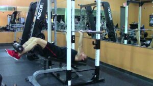
Leg Raises-7

How Testosterone Affects Fat Loss: Real Science of Low-T | Thomas DeLauer

health education on ethinylestradiol and diazepam