(USMLE topics) ACS: Symptoms, risk factors, causes, pathophysiology, diagnosis (ECG and cardiac markers), treatments. This video is available for instant download licensing here: https://www.alilamedicalmedia.com/-/galleries/narrated-videos-by-topics/cardiac-pathology/-/medias/30984947-aca5-453d-b1ae-f3a9e1f6a07d-acute-coronary-syndrome-acs-unstable-angina-nstemi-and-stem Voice by: Marty Henne ©Alila Medical Media. All rights reserved. Support us on Patreon and get early access to videos and free image downloads: patreon.com/AlilaMedicalMedia All images/videos by Alila Medical Media are for information purposes ONLY and are NOT intended to replace professional medical advice, diagnosis or treatment. Always seek the advice of a qualified healthcare provider with any questions you may have regarding a medical condition. Acute coronary syndrome, ACS, is a group of conditions that occur when blood supply to an area of the heart muscle, the myocardium, is suddenly reduced or blocked. Reduced blood supply is termed ischemia. A prolonged ischemia can lead to death of the heart tissue, known as heart attack or myocardial infarction. A typical symptom of ACS is central chest pain or discomfort that may spread to the back, jaw, left shoulder or arm. The pain may also be felt in the abdomen and can be mistaken for indigestion. Other symptoms include shortness of breath, heavy sweating, nausea, palpitations, and fainting. Women and older people often have atypical symptoms. Chest pain is not reported in about 30% of cases. About 20% of patients have mild or no symptoms. Risk factors include: aging, family history of heart diseases, smoking, diabetes, hypertension, high cholesterol, obesity, sedentary lifestyle, unhealthy diets, and psychosocial stress. The most common cause of ACS is the formation of a blood clot in an artery already narrowed by an atherosclerotic plaque. Plaques are deposits of lipids, fibrous tissue and calcium, that accumulate slowly overtime. A plaque may become inflamed and rupture, exposing substances that promote coagulation, producing a blood clot on top of the plaque. Less common causes include coronary artery embolism, coronary spasms, and coronary artery dissection. Depending on the location and the extent of obstruction, one of the following conditions may result: – Unstable angina, – Non-ST-segment elevation myocardial infarction, NSTEMI, – Or ST-segment elevation myocardial infarction, STEMI. These conditions are differentiated based on ECG findings and cardiac marker levels. Markers are substances released into the bloodstream upon death of cardiac myocytes. Cardiac troponins are the markers of choice as they become elevated early in the disease process. The amount of troponin released is proportional to the infarct size. Most patients with infarction show elevated troponin levels within 6 hours. Troponin assay is usually done on presentation and then repeated a few hours later. Unstable angina is basically ischemia without infraction. It impairs cardiac function but has not caused cell death. ECG changes are absent or transient. Cardiac troponin levels remain normal, or only slightly elevated. It should be noted, however, that this condition is clinically unstable and often precedes a myocardial infarction. NSTEMI is infraction that is limited to the inner layer of the ventricular wall. Cardiac marker levels are elevated as a result of myocardial cell death. ECG changes may include ST-segment depression, or T-wave inversion, but not ST-segment elevation. STEMI is infarction that extends the entire thickness of the myocardium. STEMI results from a complete and persistent occlusion of blood flow. ECG findings show a characteristic ST-segment elevation. There is usually a reciprocal ST depression in the electrically opposite leads. The location of infarction can be identified based on the leads with ST elevation. Cardiac markers are elevated. All patients with suspected ACS are treated promptly with antiplatelets (usually aspirin), angina medications (usually nitroglycerin) and supplemental oxygen if required. Once diagnosis is confirmed, anticoagulants, beta-blockers, ACE inhibitors, and statin are usually initiated within 24 hours unless otherwise contraindicated. For STEMI patients, emergency percutaneous coronary intervention, PCI, is the preferred procedure to restore blood flow. If PCI is not available, a fibrinolytic therapy is usually initiated at the earliest possible time. For patients with unstable angina or NSTEMI, an angiography to identify sites of blockage may be done within 24 to 48 hours if the patient is clinically stable and the case is uncomplicated. For complicated cases, angiography must be done immediately. Fibrinolytic therapies are not indicated for NSTEMI patients.
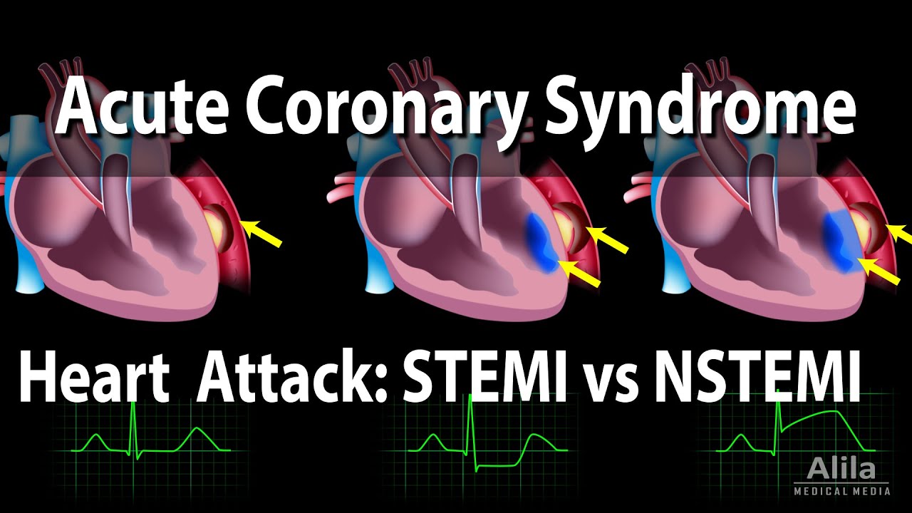
Acute Coronary Syndrome: Unstable Angina, NSTEMI and STEMI (Heart Attack), Animation
You Might Also Like
Latissimus Dorsi Bent Over Row-2
Testosterone Production

How to Grill CHICKEN BREAST | Guru Mann | Health And Fitness
Orthopedic Physiotherapy Video – 11
Gastroenterology Video – 1
Keto Diet, Keto Foods, Keto Recipes Video – 16

Donkey Kicks

skeletal system song // thrift shop parody

Cross-Cultural Psychiatry Video – 2
Step Up-2

Asanas Meaning And More Asanas Video – 5
Importance of nutrition in Human body

BCAA Kya Hai? BCAA Ke Fayde Aur Nuksan | @Fitness Fighters

Rowing Video – 2
? #1 Most Popular Prescription Drug That Causes Erectile Dysfunction & Impotence – by Dr Sam Robbins

Cross-Cultural Psychiatry Video – 3
Decline Bench Press-6

Pronunciation of Anaerobic | Definition of Anaerobic
Kidney Function Model
Neuro Surgery Video – 3

Fat Loss Weight Loss Video – 1
Omega 3 Fatty Acids के फायदे और नुकसान – Wow, Amway, Healthkart, MuscleBlaze, Healthviva Review
Keto Diet, Keto Foods, Keto Recipes Video – 29
WHEY PROTEIN or MASS GAINER? (Tips for Beginner)
THE BEAST, this is Bodybuilding!!!!
Leg Extension – Leg Curl Machine (M8XX)
Growth Hormone Deficiency

Kelli’s Quick Cool Down and Stretch – Feel Good Stretching Routine for Morning or Night

How To: Standing Overhead Tricep Rope Extensions

The Rock’s CHEST WORKOUT ROUTINE
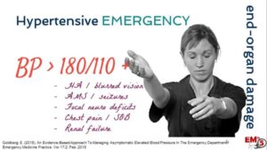
Hypertensive Emergency Treatment

Ayurved Panchakarma Video – 3

Bodybuilding biceps exercises with dumbbells — alternate hammer curl

Keto Diet, Keto Foods, Keto Recipes Video – 15
Gynecological Surgeries Video – 4

Leg Stretching Machine Types

are you ready for alli? – find out at myalli.com
The Estrogen Receptor (II): Molecular & Cellular Mechanisms
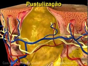
What Is Happening When Acne Forms? Does This Knowledge Help Prevent Acne?
Prime Body HCG Preparation

Donkey Kicks exercise (Beginner)